Hidden body map in mouse secondary motor cortex reframes complex movement
A study using wide-field optical imaging reports body-part-specific topography in mouse M2 during rest and movement, but human and clinical meaning remains hypothetical.

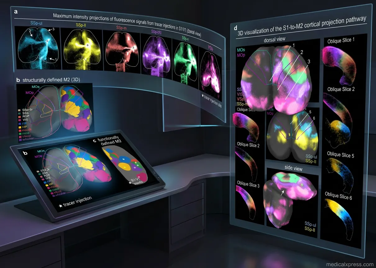

A study published in Nature Communications in 2026 reports a body-part-specific map in the secondary motor cortex, or M2, of mice. The map emerged from resting neural activity: small spots in M2 synchronized with regions of the sensory cortex that already have a long-established body map. The authors say the result challenges the view that M2 lacks such topography. [1]
The researchers used high-speed wide-field optical brain imaging while mice moved, but the report says apparently random resting activity was more revealing. When they assembled the synchronized M2 spots, they formed a body map hidden in intrinsic chatter. Two-color wide-field optical mapping distinguished the activity of corticospinal neurons, most concentrated in M1, from the activity of other neurons, confirming the map was in M2 rather than M1. [1]
The team also checked neural wiring between sensory cortex and M2, finding that synchronized subregions in each area were anatomically connected. They then verified that different mouse movements activated the M2 locations the map predicted. For example, when a mouse raised both forepaws to its mouth and licked them, each corresponding M2 point activated as those parts moved. M2 was strongly engaged when multiple body parts worked together. [1]
The result matters because coordinating several body parts at once is difficult for robots but appears effortless for animals. The study offers a structural account of how one cortical area may organize multi-part actions. Hillman, the corresponding author, expects the insights to help interpret how the brain plans and coordinates movements from moment to moment, with possible relevance to movement disorders and robot design; those are expectations, not demonstrated outcomes. [1]
Advertisement
The report also links the finding to dexterous movements that can be lost after brain injuries such as strokes. That link is a rationale for future research, not evidence that the map protects against injury or improves recovery. The study does not test rehabilitation, treatment, or patient outcomes. Its contribution is a mouse-circuit description that may guide questions about distributed cortical control. [1]
For basic neuroscience, the study revises how M2 can be viewed. The report describes M2 as chattering when the body is still and as caring more about complicated movements than simple ones. The report says these features help explain why M2 activity patterns have been so challenging to interpret. [1]
The design is a mouse imaging and behavior study, not a human clinical trial or epidemiological study. It combined wide-field optical imaging of brain activity with behavioral observation. The supplied report does not provide sample size, effect sizes, confidence intervals, or statistical details, so the precision and reproducibility of the reported map cannot be judged from the source text alone. [1]
In the resting-state analysis, the researchers identified small M2 spots whose activity patterns were synchronized to sensory cortex regions. Because sensory cortex has a long-established body map, that synchronization supplied an anchor for assigning body-part specificity. The authors then put the spots together into a map. This inference rests on spatial correlation and anatomical connection, not on stimulation-evoked movements alone. [1]
Advertisement
Two-color wide-field optical mapping was used to distinguish the activity of corticospinal neurons, which send signals down the spinal cord and are most concentrated in M1, from the activity of other neurons. That control addressed whether the apparent map actually belonged to M1. The team also checked wiring between sensory cortex and M2. Finally, behavioral tests confirmed that movement of particular body parts activated the predicted M2 subregions. [1]
Xu spent three years testing the surprising results, according to the report. That detail suggests sustained internal checking, but it is not the same as independent replication. The source does not describe preregistration, blinding, randomized sampling, or causal perturbation. Those omissions matter when judging how firmly the map should be accepted before other laboratories reproduce it. [1]
Earlier stimulation-based mapping had found that subregions of the primary motor cortex, or M1, control different body parts; stimulating one spot might cause a finger to twitch. The same approach gave less clear results for M2, leading researchers to think M2 lacked the kind of body map seen in M1. That history is the backdrop for the new resting-activity approach. [1]
Previous research had linked M2 with more complex combinations of motion, but interpreting its activity patterns in terms of body movements was especially challenging. The new study departs from stimulation-only mapping by using intrinsic resting dynamics and sensory-cortex synchronization. The report presents this as a way to expose hidden organization that movement-evoked or stimulation-based maps had not resolved. [1]
Advertisement
The source frames the work against a long philosophical question about the relationship between mind and body, and it says new tools let science revisit that question. The study does not resolve the philosophical issue. It provides mouse evidence about cortical topography and movement-related activation, which is a narrower but testable contribution. [1]
This is not an epidemiological study, so it reports no absolute risk, relative risk, odds ratio, or confidence interval for any human outcome. It cannot show that the discovered map changes a person's risk of stroke, movement disorder, or poor recovery. The appropriate inference is biological plausibility and hypothesis generation, not risk modification. Clinical claims would require human studies with defined exposures and outcomes. [1]
Absolute versus relative risk language does not apply here because the source provides no epidemiological exposure, no clinical endpoint, and no clinical risk estimate. The evidence is mechanistic: imaging, anatomical tracing, and behavioral activation in mice. Any statement that the finding lowers or raises risk in patients would exceed the supplied evidence. The study's value is in generating testable questions about cortical organization. [1]
The report also does not establish causality from the M2 map to complex movement. Activation that matches a movement is consistent with participation, but it does not prove that the subregion is necessary or sufficient. The source does not report perturbation experiments, so the causal role of M2 remains open. [1]
Advertisement
Hillman expects these insights to help interpret how the brain plans and coordinates movements from moment to moment. She suggests possible usefulness for understanding movement disorders and for building better robots. These are attributed forecasts, not results. The study did not test a movement-disorder treatment, build a robot, or demonstrate engineering benefit. [1]
Xu hopes other researchers will leverage the discovery to understand how brain activity becomes movement and how resting activity can reveal hidden wiring. That is a research agenda rather than a clinical conclusion. It points toward future work on intrinsic dynamics, connectivity, and behavior, but the supplied report does not show that such work has been done. [1]
For stroke and injury research, the map offers a candidate framework for studying dexterous movements that use more than one body part. It does not indicate that M2 is spared after injury, that it reorganizes, or that it can be targeted to improve recovery. Those remain questions for injury models and human studies, not findings of this mouse imaging study. [1]
A central unanswered question is whether a comparable body-part-specific M2 map exists in humans. The study was conducted in mice, and the report does not provide human imaging or human behavioral data. Species differences in cortical organization and movement repertoire cannot be assumed away. Human relevance is therefore a hypothesis to test. [1]
It is also unknown whether the resting-state map is stable across brain states, ages, or after injury. The report describes experiments in mice but does not supply developmental, longitudinal, or stroke-model data. Whether the map is fixed wiring, a dynamic state, or a mixture of both is not resolved by the supplied evidence. Those distinctions matter for both basic and clinical interpretation. [1]
What M2 contributes causally also remains open. Does its activity merely correlate with complex movement, or is it required for coordinating multiple body parts? The source does not report optogenetic, lesion, pharmacological, or other perturbation experiments. Without such tests, the map should be described as a topography linked to movement, not as a proven control mechanism. [1]
How robotics might use the finding is similarly speculative. Hillman expects possible robot-design relevance, but no robot was built or tested in the study. The report does not compare M2-like control with existing robotic architectures. Any engineering application is a scenario for future work, not a demonstrated outcome. [1]
The source does not report independent replication by other laboratories. It also does not report whether the finding holds in other mouse lines. Those are gaps in the evidence base, not findings of failure. [1]
A proposed next step, presented here as future work and not attributed to the source, would be independent replication. A question for that work is whether the resting-state spots remain stable and whether the behavioral activation patterns generalize. [1]
The source also does not report causal perturbation experiments, such as optogenetic, lesion, or pharmacological tests, during multi-part tasks. That absence limits causal inference. [1]
A proposed causal test, not a finding of this study, would activate or silence specific M2 subregions during multi-part tasks. The question is whether such manipulation changes coordination, which would test necessity and sufficiency. [1]
The report does not include human imaging, human behavioral data, or stroke-model data. It therefore cannot establish whether an M2-like map exists in people or changes after injury. [1]
A proposed translational question, not a result of the source, is whether an M2-like map exists in humans and whether it changes after stroke. Until such studies exist, clinical and risk implications remain questions; the bedside message is cautious. This is promising mechanistic mouse evidence, not a reason to change patient care or risk counseling. [1]
Sources
Prepared with AI assistance from the cited sources.
The Cali Reporter newsletter
The stories worth your time.
California, the country, and what comes next. Join our free newsletter for reporting that keeps you informed.
More in Health & Medicine
- Brain Energy Mismatches in Schizophrenia
A new study explores the potential role of energy bottlenecks in brain cells in explaining fragmented experiences of time, space, and self in schizophrenia.
- Brazil Reports First Mpox Death in 2026
Brazil has recorded its first mpox-related death in 2026, but public health officials are not classifying the current surge in cases as an outbreak, according to the Fiocruz Institute.
- Personalized Therapies for IBD May Target Specific Harmful Gut Bacteria
A study led by Dr. Lukas Mager and Prof. Kathy McCoy has identified two gut bacteria that promote inflammation in inflammatory bowel disease, opening the door for personalized treatments.