Early Brain Changes Linked to Alzheimer's Disease
New research suggests that Alzheimer's disease may leave detectable clues in the brain up to a decade before memory problems appear, potentially allowing for earlier treatment.

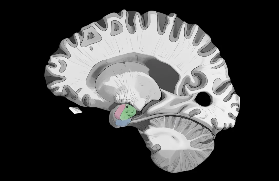
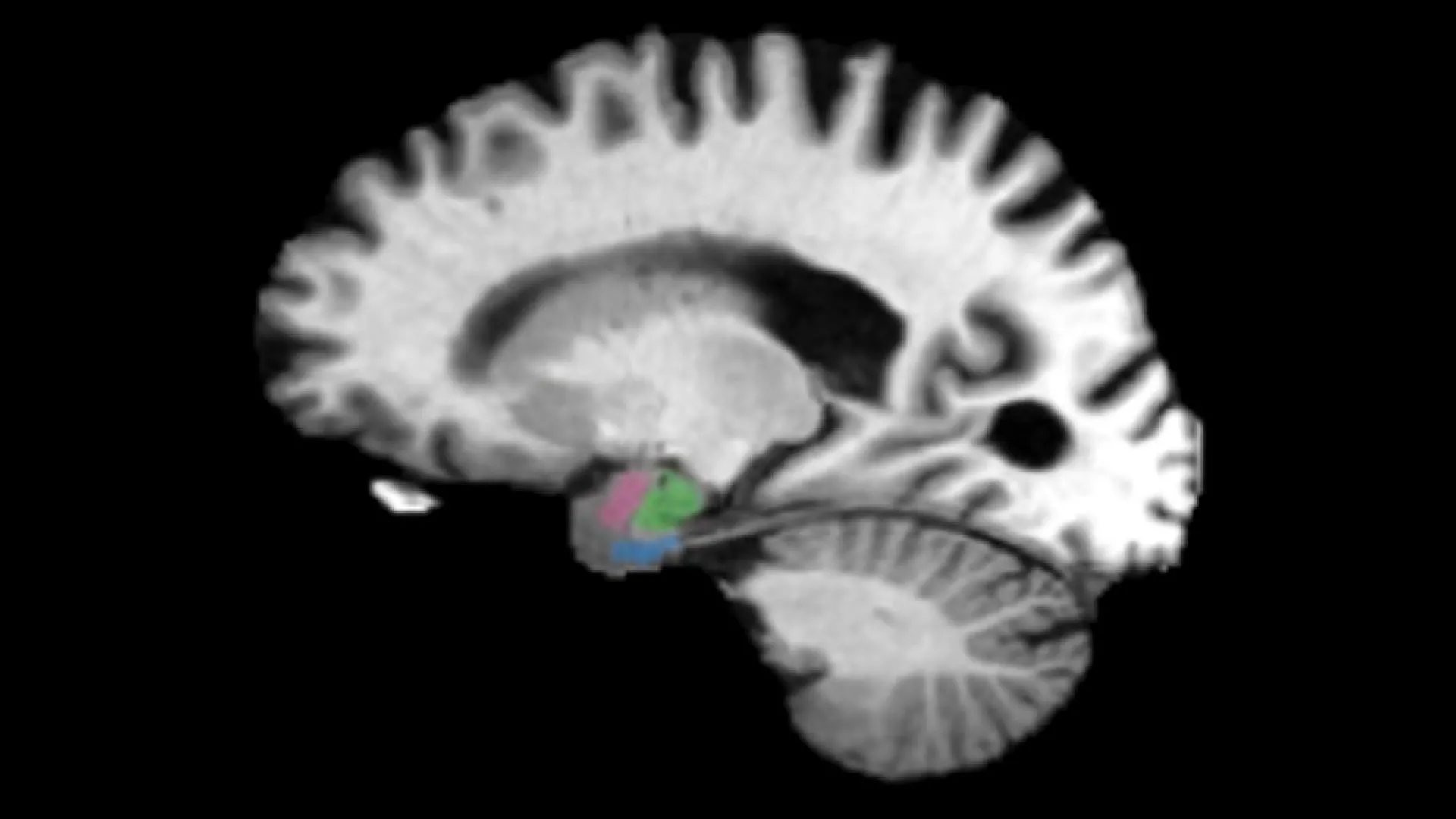
Researchers at Johns Hopkins University have identified two brain regions, the entorhinal cortex and amygdala, that show signs of deterioration earlier than the hippocampus, the area commonly examined for evidence of Alzheimer's disease (ScienceDaily Health & Medicine, 2026). These findings could help doctors recognize Alzheimer's years before symptoms develop, potentially allowing treatment to begin when it has the greatest chance of protecting the brain. The study involved examining hundreds of brain scans, revealing that these two regions begin losing tissue earlier than the hippocampus, offering new insights into the early stages of the disease. [1]
The study found that changes in the entorhinal cortex and amygdala may be detectable up to a decade before the onset of clinical symptoms, suggesting a crucial window for early intervention (ScienceDaily Health & Medicine, 2026). These changes could provide important clues about how Alzheimer's develops and how it might be detected before significant cognitive decline occurs. The researchers used magnetic resonance imaging (MRI) and positron emission tomography (PET) scans collected through two independent research projects to observe patterns of tissue loss in these brain regions. [1]
The study also found a connection between the loss of brain tissue and tau, a protein closely associated with Alzheimer's disease. Using PET imaging and digital pathology, the scientists observed that areas of gray matter shrinkage overlapped with regions showing tau-related changes (ScienceDaily Health & Medicine, 2026). This relationship strengthens the possibility that changes in the entorhinal cortex and amygdala could serve as useful indicators of early Alzheimer's disease. Researchers believe that measuring tissue loss in both regions could improve predictions of who is likely to develop cognitive decline. [1]
Researchers at Johns Hopkins University have identified two brain regions, the entorhinal cortex and amygdala, that show signs of deterioration earlier than the hippocampus, the area commonly examined for evidence of Alzheimer's disease (ScienceDaily Health & Medicine, 2026). The study found that changes in the entorhinal cortex and amygdala may be detectable up to a decade before the onset of clinical symptoms, suggesting a crucial window for early intervention (ScienceDaily Health & Medicine, 2026). These findings could allow doctors to recognize Alzheimer's years before symptoms develop, potentially enabling treatment when it has the greatest chance of protecting the brain. [1]
Advertisement
The study also found a connection between the loss of brain tissue and tau, a protein closely associated with Alzheimer's disease. Using PET imaging and digital pathology, the scientists found that areas of gray matter shrinkage overlapped with regions showing tau-related changes (ScienceDaily Health & Medicine, 2026). This relationship strengthens the possibility that changes in the entorhinal cortex and amygdala could serve as useful indicators of early Alzheimer's disease. The entorhinal cortex helps retrieve previously stored memories, while the amygdala contributes to emotional memory, suggesting that examining these structures together could reveal signs of Alzheimer's that might be less apparent when attention is focused primarily on the hippocampus. [1]
The researchers examined hundreds of magnetic resonance imaging (MRI) and positron emission tomography (PET) scans collected through two independent research projects: the Alzheimer's Disease Neuroimaging Initiative and the Biomarkers of Cognitive Decline Among Normal Individuals study (ScienceDaily Health & Medicine, 2026). In both groups, the researchers observed a consistent pattern, with the entorhinal cortex and amygdala showing greater relative tissue loss during the early stages of Alzheimer's than the hippocampus itself. This distinction matters because the hippocampus plays a central role in memory and has traditionally been a major focus when doctors look for Alzheimer's-related brain damage. [1]
Researchers at Johns Hopkins University have identified two brain regions, the entorhinal cortex and amygdala, that show signs of deterioration earlier than the hippocampus, the area commonly examined for evidence of Alzheimer's disease (ScienceDaily Health & Medicine, 2026). The study found that changes in the entorhinal cortex and amygdala may be detectable up to a decade before the onset of clinical symptoms, suggesting a crucial window for early intervention (ScienceDaily Health & Medicine, 2026). These findings could help doctors recognize Alzheimer's years before symptoms develop, potentially allowing treatment to begin when it has the greatest chance of protecting the brain. [1]
The researchers examined hundreds of magnetic resonance imaging (MRI) and positron emission tomography (PET) scans collected through two independent research projects: the Alzheimer's Disease Neuroimaging Initiative and the Biomarkers of Cognitive Decline Among Normal Individuals study (ScienceDaily Health & Medicine, 2026). The scans followed people at different stages of Alzheimer's disease over time, including individuals without symptoms, those experiencing cognitive impairment, and patients who ultimately died from the disease. In both groups, the researchers observed a consistent pattern: the entorhinal cortex and amygdala showed greater relative tissue loss during the early stages of Alzheimer's than the hippocampus itself. [1]
Advertisement
The researchers also found an important connection between the loss of brain tissue and tau, a protein closely associated with Alzheimer's disease (ScienceDaily Health & Medicine, 2026). Using PET imaging and digital pathology, the scientists found that areas of gray matter shrinkage overlapped with regions showing tau-related changes. This relationship strengthens the possibility that changes in the entorhinal cortex and amygdala could serve as useful indicators of early Alzheimer's disease. [1]
Researchers at Johns Hopkins University have identified two brain regions, the entorhinal cortex and amygdala, that show signs of deterioration earlier than the hippocampus, the area commonly examined for evidence of Alzheimer's disease (ScienceDaily Health & Medicine, 2026). The study found that changes in these regions may be detectable up to a decade before the onset of clinical symptoms, suggesting a crucial window for early intervention (ScienceDaily Health & Medicine, 2026). These findings were based on the analysis of hundreds of brain scans collected through two independent research projects: the Alzheimer's Disease Neuroimaging Initiative and the Biomarkers of Cognitive Decline Among Normal Individuals study (ScienceDaily Health & Medicine, 2026). [1]
The study also found a connection between the loss of brain tissue and tau, a protein closely associated with Alzheimer's disease (ScienceDaily Health & Medicine, 2026). Using PET imaging and digital pathology, the scientists found that areas of gray matter shrinkage overlapped with regions showing tau-related changes (ScienceDaily Health & Medicine, 2026). This relationship strengthens the possibility that changes in the entorhinal cortex and amygdala could serve as useful indicators of early Alzheimer's disease (ScienceDaily Health & Medicine, 2026). [1]
The researchers examined hundreds of magnetic resonance imaging (MRI) and positron emission tomography (PET) scans collected through two independent research projects: the Alzheimer's Disease Neuroimaging Initiative and the Biomarkers of Cognitive Decline Among Normal Individuals study (ScienceDaily Health & Medicine, 2026). The scans followed people at different stages of Alzheimer's disease over time, including individuals without symptoms, those experiencing cognitive impairment, and patients who ultimately died from the disease (ScienceDaily Health & Medicine, 2026). In both groups, the researchers observed a consistent pattern of greater relative tissue loss in the entorhinal cortex and amygdala during the early stages of Alzheimer's than in the hippocampus (ScienceDaily Health & Medicine, 2026). [1]
Advertisement
The Cali Reporter newsletter
The stories worth your time.
California, the country, and what comes next. Join our free newsletter for reporting that keeps you informed.
More in Health & Medicine
- Brain Energy Mismatches in Schizophrenia
A new study explores the potential role of energy bottlenecks in brain cells in explaining fragmented experiences of time, space, and self in schizophrenia.
- Brazil Reports First Mpox Death in 2026
Brazil has recorded its first mpox-related death in 2026, but public health officials are not classifying the current surge in cases as an outbreak, according to the Fiocruz Institute.
- Personalized Therapies for IBD May Target Specific Harmful Gut Bacteria
A study led by Dr. Lukas Mager and Prof. Kathy McCoy has identified two gut bacteria that promote inflammation in inflammatory bowel disease, opening the door for personalized treatments.